Live Imaging System
Formerly known as the Live Cell Imaging platform
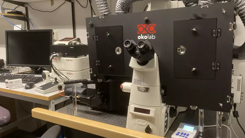
Description
Live imaging is an important tool for studying the molecular and cellular functions of neuronal and non-neuronal cells and tissue. It has a broad range of applications, such as exploring disease mechanisms, assisting with clinical diagnosis and development of new treatments. This system gives MultiPark scientists an opportunity to study single molecule dynamics in high resolution or to track cell migration and cell differentiation long-term.
The system that MultiPark has under the Microscopy Platform is a Nikon Eclipse Ti equipped with a controllable enclosure. The microscope is fully motorized and can detect signal via an EMCCD camera or X-light V2 spinning disk confocal unit. This unit is suitable for imaging with fluorescence labeling, such as DAPI, CFP, GFP, YFP, mCherry and Cy5.
Using the system
The system is located in BMC A1024 and is booked via Outlook calendar. For more information, please contact Megg (megg [dot] garcia-ryde [at] med [dot] lu [dot] se) or Gunnar (gunnar [dot] gouras [at] med [dot] lu [dot] se).
Contact for training/access

Megg Garcia-Ryde
Research engineer
Email: megg [dot] garcia-ryde [at] med [dot] lu [dot] se
Office: BMC B1128b
Responsible PI
Gunnar Gouras
Professor
Email: gunnar [dot] gouras [at] med [dot] lu [dot] se
Office: BMC B1123c